Mammography
Successful treatment of breast cancer depends on early diagnosis
Mammography plays a vital role in early diagnosis. By examining the digital images from your mammogram, your doctor can see changes in your breast tissues up to two years before either you or your doctor can feel them. According to the Food and Drug Administration (FDA), mammography can detect 85 to 90 percent of breast cancers in women over 50. Current guidelines from the American Medical Association (AMA) and the American College of Radiology (ACR) recommend that beginning at age 40 women get annual mammograms.
Mammography is a specific type of imaging that uses a low-dose x-ray system. Thanks to a collaboration with Bloomington Hospital and the Bloomington Hospital Foundation, SIRA now offers high-contrast, high-resolution digital imaging for examination of the breasts. Using digital imaging allows your doctor to more easily compare, manipulate and magnify the images for a better view. Digital images of tissues near the skin and breastbone are much more clear than film, and they provide much better scans for women with dense breast tissues, using less radiation than analog mammography. Digital technology also takes less of your time: the images seldom need to be retaken, but if they do, we can take care of it right away.
There are two types of mammograms. A diagnostic mammogram is ordered to diagnose problems such as lumps, thickening, inverted nipple, or discharge. A screening mammogram is a “routine” exam. This is done when you have no known problems with your breasts.
How to schedule a mammogram
If you have not already scheduled a mammogram through your primary care physician or OB/GYN, you can call SIRA at 812-333-7676 and select option 1. An assistant there will take your call and walk you through the steps to get a mammogram appointment. If you do not have a primary care physician or OB/GYN to place the order for your mammogram, you will be given the contact information for one of several local physician referral sites including IU Health providers for referrals or call 800-248-1199, the Monroe Owen County Medical Find a Physician or call 812-332-4033, Planned Parenthood in Bloomington or Bedford (812-336-0219 or 812-279-3527), or Volunteers in Medicine (812-353-3533).
How to prepare for your mammogram
Before scheduling your mammogram, discuss any new findings or problems in your breasts with your doctor. Also, inform your doctor of any prior surgeries, hormone use, and family or personal history of breast cancer.
Do not schedule your mammogram for the week before your period if your breasts are usually tender during this time. The best time is one week after your period ends. Always inform your doctor or x-ray technologist if there is any possibility that you are pregnant.
What to expect at your appointment
Do not wear deodorant, powder, perfumes or lotion on the day of the exam. These can appear on the x-ray film as calcium spots.
Describe any breast symptoms or problems to the technologist performing the exam.
If you have them, bring along copies of prior mammograms and give them to the technologist when you arrive for your exam.
Before the examination, you will be asked to remove all jewelry and clothing above the waist and you will be given a gown that opens in the front.
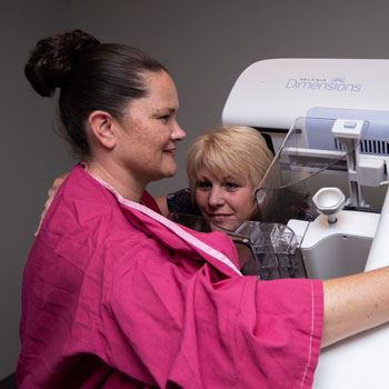
What happens during your mammogram?
A specially trained and certified female mammography technologist will call you into the exam room. Because of the radiation, no one else is permitted into the room with you while the exam is done. The technologist will review your history with you and make notes for the radiologist concerning scars, moles, lumps, and comments you have concerning your breasts.
During your mammogram, your technologist will position you carefully. The tech will use the special paddles on the machine to compress your breast. It is necessary that your breast be compressed so that the breast tissue thickness is evened out and spread as much as possible. If there is a lesion in the underlying tissues, this compression will enable your doctor to better see it. If you experience significant discomfort, be sure to tell the technologist.
You will maintain this position for just a few seconds while the technologist does the exam.
You will have to hold very still for a few seconds while the unit works. Other than the momentary discomfort that you may feel from the compression, you will not experience any pain.
The technologist will reposition you for a second view of the same breast.
Your mammogram will consist of at least two different positions for each breast, perhaps more if we have to to get a good result.
What to expect after the exam
If you are having a screening (routine) mammogram, the technologist will review your images before you go to determine whether the images are clear enough or whether they need to be retaken. The radiologist will read your exam after you leave and send a report to your doctor. Because you leave before the radiologist sees your images, there is a small chance that you may have to return for additional pictures to be taken.
If you are having a diagnostic mammogram, the technologist will process the images and a radiologist will review them while you wait. Sometimes the radiologist may need additional tests, such as ultrasound, to make a final report for your doctor. The radiologist will talk with you before you leave and give you your results.
One of our radiologists will analyze the images of your breasts, dictate a report on any findings, and suggest a diagnosis. A report will be sent to your referring physician.
After reviewing the information from our radiologists, your healthcare provider may schedule you to return to SIRA for further testing or biopsies.
False positive mammograms
Between 5 and 10 percent of mammogram results are abnormal and require more testing (more mammograms, fine needle aspiration, ultrasound, or biopsy), and most of the follow-up tests confirm that no cancer was present. It is estimated that a woman who has yearly mammograms between ages 40 and 49 would have about a 30 percent chance of having a falsely-positive mammogram at some point in that decade, and about a 7 to 8 percent chance of having a breast biopsy within the 10-year period. The estimate for false-positive mammograms is about 25% for women ages 50 or older.
The risks of mammography
Mammography is an x-ray imaging process. The exposure to radiation received from two digital mammographic views is equivalent to less than six months of natural exposure (including cosmic radiation from outer space, radiation from the soil and buildings, and natural isotopes in our own bodies). This amount is believed to be safe.
We take special care during any x-ray examination to assure maximum safety for you by shielding you with a lead apron where appropriate. Inform your doctor and your technologist if there is any possibility that you are pregnant.
Mammograms cannot provide you 100% protection
As explained in the first paragraph of this page, mammography, like any other medical test, can sometimes miss a problem. To more closely insure your good breast health, it is important that you perform breast self-exams monthly and visit your primary care physician for manual exams annually. The combination of these safeguards is the best advice medicine can give you.